- Home
- Knowledge & Research Center
- Scientific Articles
- Laser-Assisted Preprosthetic Surgery with Waterlase – Soft Tissue and Bone Preparation
Laser-Assisted Preprosthetic Surgery with Waterlase – Soft Tissue and Bone Preparation
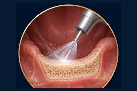
Scientific and clinical overview of laser-assisted preprosthetic surgery with Waterlase. Gingival and mucosal reshaping, bone contouring, and functional preparation of the prosthetic site. Includes a clinical case with postoperative photographic documentation.
Waterlase-Assisted Preprosthetic Surgery
Gingival, mucosal and osseous preparation for functional prosthetic rehabilitation.
Abstract
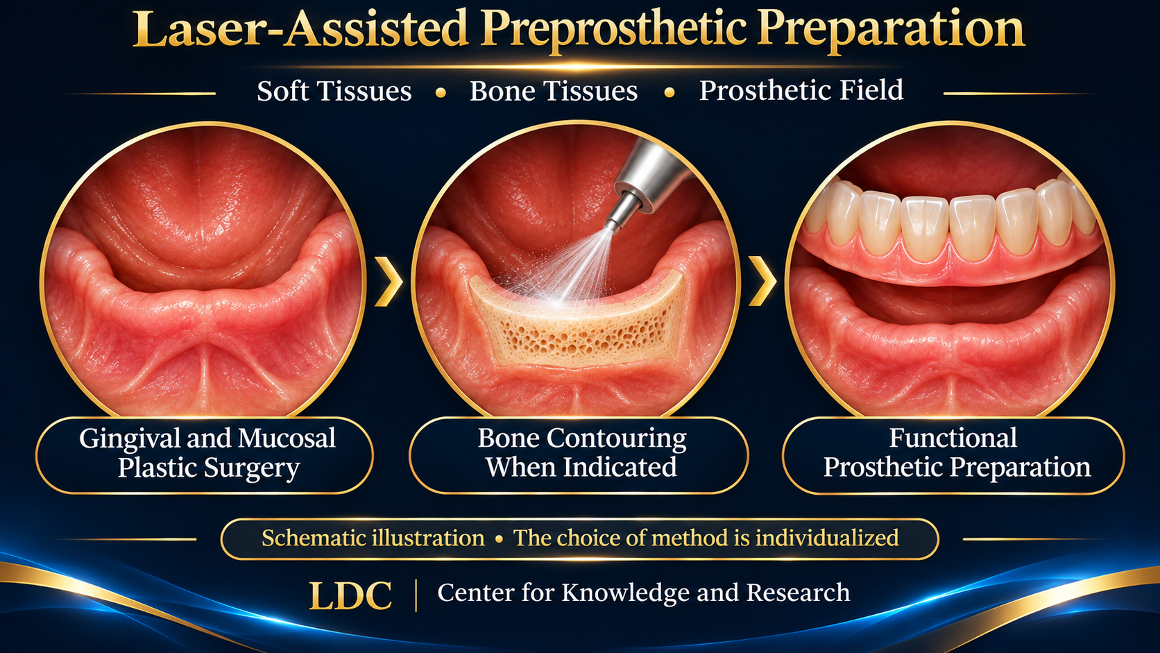
Successful prosthetic rehabilitation depends on both the prosthesis and the quality of its supporting tissues. In selected cases, Waterlase (Er,Cr:YSGG, 2780 nm, with an air-water spray) may be used for soft-tissue contouring and oral osseous reshaping. Preprosthetic surgery aims to optimize the prosthetic field as part of an individualized treatment plan; laser-based techniques do not universally replace conventional surgery.
Why preprosthetic preparation matters
Irregular alveolar ridges, shallow vestibules, high muscle insertions and mobile or fibrotic mucosal tissue may compromise the retention, stability and comfort of removable prostheses. Assessment of anatomy, tissue health and prosthetic goals determines whether the patient benefits from conservative treatment, soft-tissue modification, osseous recontouring or a combined approach.
Three interrelated clinical directions
Gingival contouring: selective reshaping of soft-tissue margins when required by the prosthetic plan and tissue biology.
Mucosal surgery and vestibular optimization: management of mobile tissue, frenal attachments and vestibular depth where clinically indicated.
Osseous contouring / alveoloplasty: conservative correction of irregular bone contours, with attention to cooling, thermal safety and preservation of supporting tissues.
Waterlase tissue interaction
An Er,Cr:YSGG device emits at 2780 nm. Laser energy interacts with tissue water and mineralized tissue to produce ablation under appropriate conditions. Air-water spray aids cooling and debris removal. Clinical safety and precision depend on the operating mode, handpiece and tip, settings, water delivery, anatomy and operator training. Thermal injury remains possible if the technique is inappropriate.
Current evidence and limitations
A 2026 study involving 36 completely edentulous patients compared Nd:YAG, diode and Er:YAG laser vestibuloplasty with conventional scalpel surgery, reporting differences in postoperative symptoms and maintenance of vestibular depth. It did NOT specifically study Waterlase (Er,Cr:YSGG). The small sample, single-center setting and different devices limit generalization. A 2024 case report described Er,Cr:YSGG-assisted alveoloplasty and soft-tissue surgery before prosthetic rehabilitation. These reports support feasibility in selected indications but not universal claims of faster healing or better long-term prosthetic outcomes.
Laser Dental Clinic: clinical perspective
At Laser Dental Clinic, water-assisted laser procedures are used for selected gingival and mucosal corrections and, where indicated, osseous recontouring within comprehensive preprosthetic planning. This particular documented case concerns soft-tissue treatment; the photographs alone do not demonstrate that an osseous procedure was performed. The intended outcome is a suitable prosthetic-bearing field, with subsequent healing and prosthetic adaptation documented at follow-up.
Clinical case: soft-tissue preparation before complete denture rehabilitation
According to the treating clinician, the case concerns preprosthetic preparation before complete denture rehabilitation. The preoperative findings included thickened mobile mucosa with multiple wart-like projections in the planned prosthetic-bearing field. A Waterlase-assisted soft-tissue procedure was performed. Both available photographs depict the site AFTER the intervention; they are not before-and-after comparisons, and the precise postoperative timepoint has not been established. “Wart-like” is a morphological description, not evidence of viral etiology or a confirmed histopathological diagnosis. The images do not establish complete healing, osseous surgery in this particular case, or the final prosthetic outcome.
Diagnostic note: Persistent, atypical or suspicious papillary/verrucous oral lesions warrant appropriate biopsy and histopathological assessment. The pathology status was not supplied for this editorial draft; this does not imply that no diagnostic procedure occurred. [6]
What would complete the clinical case?
Preoperative findings, location and diagnosis of hyperplastic mucosa and wart-like projections; pathology information if obtained.
Technique, Waterlase model and clinically relevant operating parameters.
Dates of postoperative photographs and defined follow-up findings.
Photographs of the definitive complete denture rehabilitation; confirmation of the cast-metal prosthetic construction described by the clinician, prosthetic adaptation and functional assessment.
Specific patient permission for public use and de-identification review.
Conclusion
Laser-assisted preprosthetic surgery can support selected cases when guided by sound anatomical assessment and prosthetic planning. Benefits must be documented through appropriate outcome measures. Technology is a tool within individualized care, not a guarantee of painless treatment, accelerated healing or permanent success.
Scientific references
1. Nica DF et al. Preprosthetic vestibuloplasty performed by three different types of lasers versus conventional scalpel. Front Oral Health. 2026;7:1936637. doi:10.3389/froh.2026.1936637.
2. Emam NS, Abdelhamid AM, Segaan LG, Khamis MM. Pre-prosthetic atraumatic laser-assisted mandibular rehabilitation: Er,Cr:YSGG 2780 nm for alveoloplasty and mucocele removal. Laser Therapy. 2024;31:393. doi:10.4081/ltj.2024.393.
3. Lim KO, Lee WP. Technical Note on Vestibuloplasty around Dental Implants Using Erbium YAG Laser-Assisted Periosteal Fenestration. Medicina. 2023;59(10):1884. doi:10.3390/medicina59101884.
4. Blagova B et al. Histomorphological alterations in human bone tissue following in vivo osteotomy: conventional drilling, piezosurgery and Er:YAG laser ablation. Oral Maxillofac Surg. 2026. doi:10.1007/s10006-026-01612-x.
5. Albahdly NM, Hameedi AD. Ex vivo histological comparison between Er,Cr:YSGG laser and slow-speed handpiece in bone cutting. Wiad Lek. 2026;79(9):2102–2112. doi:10.36740/WLek/230884.
6. American Dental Association. Oral Cancer Guideline (2026 Update): biopsy remains the diagnostic standard for suspicious oral mucosal lesions. https://www.ada.org/resources/research/science/evidence-based-dental-research/oral-cancer-guideline
CLINICAL PHOTO DOCUMENTATION
Postoperative images