- Home
- Clinical Disciplines
- Cariology and Restorative Treatment
Cariology and Restorative Treatment
Minimally Invasive Treatment and Biological Restoration of the Tooth
Dental caries is a multifactorial, biofilm-mediated process involving interactions between the oral microbial ecology, dietary factors, salivary properties, the condition of the hard dental tissues, and individual risk factors
A healthy tooth means more than the absence of pain or a visible “cavity”. Caries can begin without obvious signs, while loss of hard dental tissues may also have non-carious causes.
At Laser Dental Clinic, our aim is to detect changes as early as possible, determine their cause and true extent, and preserve as much healthy natural tooth structure and pulp vitality as possible.
Treatment is not limited to removing caries and placing a “filling”. We restore the tooth’s anatomy, biology and function and monitor the result over time.
Detecting the problem as early as possible
Diagnosis begins with a careful clinical examination and assessment of the individual caries risk.
When necessary, we use DIAGNOcam to detect and monitor carious changes without X-ray exposure, together with intraoral radiographs according to the specific diagnostic indications.
Bitewing radiography supports the detection of hidden approximal caries between neighbouring teeth, assessment of lesion depth and evaluation of existing restorations.
When assessment of the root and surrounding tissues is required, a periapical intraoral radiograph is performed, including for the detection and follow-up of changes in the periapical region.
For deep carious lesions and suspected pulpal involvement, diagnostics are supplemented by assessment of pulp vitality, including EOD, according to clinical indications.
A small spot does not always mean a small lesion
Caries does not always begin as an obvious cavity and may remain painless for a long time.
We therefore pay particular attention to pinpoint and fissure lesions. A small visible opening in the enamel does not always reflect the true extent of the process beneath the surface and should not be overlooked.
A white-spot carious lesion represents an early change in enamel mineralization. Not every white spot is active caries, so we assess its activity, surface characteristics and development over time.
When the enamel surface remains intact, preventive and remineralizing approaches may support control of the demineralization process.
However, when the enamel structure is already irreversibly disrupted, cavitated or structurally defective, the lost enamel architecture cannot simply be “rebuilt” by remineralization. Treatment is then determined by the actual condition of the tissues.
Distinguishing carious from non-carious lesions
Not every loss of hard dental tissue is caused by caries, and not every carious lesion develops on an intact natural tooth.
In addition to primary carious lesions, we assess secondary carious defects around or beneath existing restorations, particularly where marginal adaptation is compromised, restorative material has changed, or conditions favour bacterial biofilm retention.
Special attention is given to carious changes beneath crowns and other prosthetic restorations. These may remain hidden from the patient and do not always produce early symptoms. When suspected, the area is assessed clinically and with appropriate imaging and additional diagnostic methods, while the decision to preserve, repair or replace the restoration is made individually.
In cases of gingival recession, dehiscence and exposed root surfaces, we assess the risk and presence of root caries. Root surfaces differ structurally from enamel and require timely detection, an individualized therapeutic approach and follow-up.
We separately assess non-carious lesions – abrasion, attrition, erosion, cervical defects, cracks and other structural changes related to mechanical, chemical, occlusal, thermal or combined influences.
Carious and non-carious changes may overlap and coexist. Before restoration, we therefore determine the cause, activity and depth of the defect, the condition of existing restorations or prosthetic constructions, and the true residual volume of healthy hard dental tissues.
What matters is not only the defect, but how much healthy tissue remains
Treatment planning includes assessment of the volume of lost hard dental tissues and the quality of the remaining tooth structure.
We consider which surfaces are involved, how much sound enamel and dentine remain, whether walls or cusps are weakened, whether cracks are present, the condition of the pulp, and how the tooth carries functional load.
For a limited defect, we aim for a maximally conservative direct adhesive restoration.
With greater tissue loss, the restoration must replace not only missing tissue but also restore anatomical form, contacts with neighbouring teeth, occlusal morphology and functional stability.
When the remaining tooth structure no longer allows a predictable direct restoration, the need for a broader protective or indirect restoration and, when indicated, subsequent prosthetic treatment is assessed.
Minimally invasive and laser-assisted treatment
When enamel or dentine is irreversibly damaged, our aim is to remove what is necessary rather than the maximum amount of tooth structure.
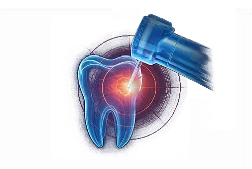
When indicated, we use a water laser for precise and minimally invasive treatment of affected hard dental tissues.
This approach may also be used for selected fissure and structural defects. When enamel cracks are present, we assess their depth and extent, associated caries, pulp status and occlusal loading.
Our goal is not to remove more tooth structure, but to determine which tissue can be preserved and which can no longer provide biological and functional stability.
When caries approaches the pulp
Deep caries does not automatically mean that the tooth must be devitalized.
In deep carious lesions, we assess the condition of the pulp and the possibility of preserving its vitality.
When the pulp retains its healing potential and the clinical conditions allow, the aim is to preserve as much vital pulp tissue as possible.
After careful removal of infected tissues, laser-assisted and biologically oriented approaches may be used together with appropriate bioactive calcium-silicate materials to create conditions for preservation of vitality and support the pulp’s natural reparative potential.
When specifically indicated, this may include vital pulp therapy with partial or complete removal of affected coronal pulp while aiming to preserve vital radicular pulp, followed by an appropriate bioactive material, restoration and follow-up.
When the pulp can no longer be biologically preserved, treatment continues within the Endodontics clinical discipline.
Creating the conditions for a precise restoration
The quality of an adhesive restoration also depends on reliable control of the operative field.
When the clinical situation permits, we use OptraDam to isolate the working area from moisture and support a clean, dry and controlled operative field.
For patients with a pronounced gag reflex, limited mouth opening or trismus, we use an individualized adaptation approach according to the cause and clinical condition.
In suitable patients, our clinical practice may include Taopatch positioned in individually selected areas as an adjunctive approach. It may be combined with gradual adaptation to procedures and individually selected exercises for tongue control and position, swallowing and related musculature.
The aim is improved patient comfort and adaptation, together with a more stable operative field for precise clinical work.
Restoring anatomy and function
After treatment of the damaged tissues, the tooth is restored using an appropriate adhesive technique and a light-cured composite material – flowable or with suitable consistency and mechanical properties according to the individual clinical situation.
Our aim is not simply to “fill the defect”.
We restore the tooth’s anatomical form, contact points with neighbouring teeth, occlusal morphology and functional relationships.
This is followed by careful finishing and polishing, control of contact points, and verification and adjustment of the occlusion.
As a final step, an appropriate protective surface coating/material is applied in accordance with the restorative system used. The aim is to support protection of the restored surface and the longevity of the result.
A restoration does not end on the day it is placed
Restorations are subsequently monitored within the patient’s individualized Maintenance Programmes.
At follow-up visits, we assess integrity, surface condition, marginal adaptation, contact points, occlusion and functional loading. We also monitor areas around existing restorations and crowns, as well as exposed root surfaces, for early detection of secondary or root caries.
When a limited defect is identified and the clinical conditions permit, we prefer minimally invasive correction or repair of the existing restoration rather than its automatic complete removal and replacement.
In this way, we aim to extend the functional life of the restoration while preserving as much healthy natural tooth structure as possible.
Our goal
Cariology and restorative treatment are not simply “caries treatment and placement of a filling”, but a structured process:
Early diagnosis → distinction between carious and non-carious lesions → assessment of tissue loss and residual hard dental tissue volume → minimally invasive treatment → preservation of pulp vitality whenever possible → precise adhesive restoration → functional and occlusal control → protection → long-term follow-up and repair when necessary.
The goal is maximum preservation of natural dental tissues and tooth vitality, together with restoration of biology, anatomy, aesthetics and function for as long as possible.